Translate this Page

Thursday, September 25, 2014

Branchial Cleft Cyst

Every so often, a pediatrician will encounter a child with a neck mass. The differential diagnosis for a neck mass is rather large, and some causes are quite frightening, but one of the best case scenarios is a simple branchial cleft anomaly. These are the culprit in 30% of reported neck masses.


Branchial cleft anomalies ocur due to a glitch in embryonic fetal development. Human fetuses begin with gills, much life fish, but they evolve into more complex structures such as facial muscles and bones, neck cartilage and major cardiac blood vessels. The anomalies appear as as cysts (no opening), fistulas (both internal and external opening) or sinuses (either internal or external openings).
 
There are four branchial cleft anomalies. Anomalies of the first arch manifest as external openings and affect either the parotid gland (cheek), ear or jaw. Anomalies of the second arch are usually cysts (95%) with no openings and occur at the level of the neck.  Anomalies of the third and fourth arches are very rare, occur on the left side (80%), and open internally at an area of the mouth called the piriform recess (90% of cases).

All branchial cleft masses can become complicated by infection, and may grow large enough to cause trouble with swallowing or breathing. They are, however, benign and can easily be removed operatively by a surgeon.

It is important for the pediatrician to consider other common causes of neck masses, such as thyroglossal duct cysts or thyroid nodules. More importantly, emergent causes of neck mass such as lymphadenitis or even malignancy should be ruled out before proceeding with treatment.

Reference
Branchial cleft anomalies. Dynamed database. Updated July 24, 2012. Accessed September 24, 2014.
 

Tuesday, September 23, 2014

Ear Infection in Children

Ear infections are one of the most common disease processes seen by pediatricians. It is estimated that up to 85% of children suffer from at least one episode of ear infection before the age of 3.


The typical age range during which ear infections are seen is from 6 to 15 months old. Symptoms range from mild pain to hearing loss and brain abscess. There are three areas that become infected in the ear, and to understand this requires some knowledge of the anatomy:


The external or outer ear is where otitis externa (OE), also known as swimmer's ear, occurs. This is  is the only infection that causes pain with manipulation of the ear. The outer ear will usually be red and tender the the touch. The most common cause of this is a bacteria called pseudomonas, which thrives in wet conditions. The treatement for this, therefore, is over the counter ear drops that dry out the ear canal and make the area uninhabitable for the bacteria.

The middle ear is behind the eardrum or tympanic membrane. It is the space that houses the ear ossicles, the bones which transmit sound from the eardrum to the cochlea of the inner ear. This area is connected to the back of the nose (pharynx) via a hollow tube known as the eustachian tube.  An infection of this space is known as otitis media (OM). There are different types of OM, depending on the length of time it has persisted and whether or not fluid (effusion) is present.

Acute otitis media (AOM) typically occurs in <2 years old, usually caused by viruses such as RSV, cytomegalovirus (CMV) and Haemophilus influenzae (Hflu) but can also be due to bacteria. Mucosal congestion caused by the upper respiratory infection (URI) is aspirated up into the middle ear where inflammation occurs.
Treatment: If younger than 2 years old, an antibiotic such as amoxicillin will be prescribed. If older than 2 years old, doctors may choose to wait 48 hours to see if symptoms improve on their own before deciding to add an antibiotic. This is because studies have shown spontaneous resolution (without treatment) in up to 80% of this age group in 2-14 days.

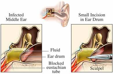
Otitis media with effusion (OME) occurs when bacteria gets into the middle ear and fills it with pus. In growing children less than 7, the eustachian tube is often short and less vertical, so pus cannot drain properly and begins to back up. This growing pocket of fluid causes a large amount of pressure and pain to the child. The doctor will use a pneumatic otoscope to blow a burst of air at this ear drum. Normally the ear drum will move but in the case of OME, no movement occurs. Tympanometry may also be used to determine this. Long term OME (>3 episodes in 6 months or >4 episdoes in 12 months) may lead to hearing and language problems, so treatment is often indicated.
Treatment: myringotomy (hole is cut into the ear drum) with tympanostomy tube insertion is surgically performed. This allows fluid to freely drain out of the ear instead of backing up. It also alows antibiotic drops to be placed in the ear, which move through the tube and into the middle ear space to treat bacterial infections. Parents are instructed to apply the drops for 10 days whenever they see pus draining from the ears, which allows them to treat infections at home.


Reference
American Academy of Family Physicians, American Academy of Otolaryngology-Head and Neck Surgery, American Academy of Pediatrics Subcommittee on Otitis Media with Effusion. Otitis media with effusion. Pediatrics 2004; 113(5): 1412-29.

Thursday, July 10, 2014

Tay Sachs Disease

Tay Sachs disease is a type of degenerating neurological disease caused by a deficiency of the enzyme protein hexosaminidase A. Because the enzyme is deficient, a lipid molecule called GM2 ganglioside builds up inside the lysosomes of cells. It's for this reason that doctors categorize it as a type of lysosomal storage disease.


The disease occurs when parents that are BOTH carriers of the genetic mutation have a baby. The most common carriers are Ashkenazi Jews, French Canadians and Louisiana Cajuns (1 in every 27), though carrier frequencies are also high in Irish Americans (1 in 50) and the general population (1 in 250). 


The disease typically occurs acutely in infancy, but there are three documented presentations: acute, subacute and late onset. 

The classic presentation, the acute (infantile) form, involves progressive motor weakness with loss of milestones and myoclonic jerking beginning at 3 months of age. 98% of patients develop seizures, and death typically occurs within the first few years of life. The severity is due a complete absence of the hexosaminidase A enzyme.

The subacute (juvenile) form will present as late as 9 years old, and is characterized by seizures, choreiform movements, speech problems and incontinence.  These children will typically develop dementia and won't live past the teenage years. 

Late onset Tay Sachs (LOTS, adult) cases have been reported in older patients, and usually involve subtle disturbances in speech, mood and psychological state. 

Pediatricians should suspect Tay Sachs Disease in any Jewish patient or patient of Jewish descent with delayed milestones and myoclonic jerks before the age of 1. Close inspection of the eye will typically reveal a cherry-red colored spot on the macula of the retina, which is only present in a few childhood diseases (Krabbe, Canavan, Alexander, and Gaucher diseases). A blood test will officially confirm the diagnosis. 


The differential diagnosis of neurologic progression in pediatric patients involves neurodegenerative disorders (spinal muscular atrophy/SMA, amyotrophic lateral sclerosis/ALS, Friedreich ataxia/FA and leukodystrophy)

Management of the disease involves maintaining nutrition, respiratory function and motor function while minimizing infectious diseases and seizures. Later forms benefit from both speech and physical therapies. All patients and their families should be referred to a certified genetic counselor for counseling. 

The prognosis for Tay Sachs disease is very poor. One retrospective study followed 97 infants diagnosed with Tay Sachs disease and noted that death occurs before the age of 5 in nearly all patients. Milestones such as head control, transferring objects from hand to hand and sitting without suport were acheived by the babies in over half the cases, but all lost their abilities by 1 year of age. The median lifespan of the infants was only 47 months.


Reference
Tay Sachs Disease. Dynamed Database. Updated May 12 2014. Accessed July 10, 2014. 

Monday, July 7, 2014

Your Child's Development: Pediatric Milestones

"Is my child developing normally?" 

This question is almost guaranteed during nearly all well child encounters in the pediatric office setting. Parents, rightfully so, are commonly concerned about their child's development and need a small amount of  reassurance from their professional caregiver. What are the guidelines that all pediatricians follow? 


The pediatrician references a set list of known guidelines when evaluating the development of a child. He or she often does this in three ways: using an Ages and Stages Questionnaire (sensitivity 75%, specificity 81%) filled out by the parent, asking the parent specific questions about the child and physically examining or interviewing the child. 


Below is the list of developmental milestones. It's important to remember two things:
1) The milestones are simply a guide: physicians use it to help determine if a child has fallen below normal when compared to other age-matched children. It is the job of the pediatrician to recognize a child that has fallen behind in one area and to offer support or resources to help him or her catch up (i.e, the Early Steps program).

2) All children develop differently. One child may meet all milestones on time while another may be persistently behind in one area. It's important to always be patient with (and supportive of) your child.

Pediatric milestones by age:

1 month
Vision: regards face, fixes transiently
Social: smiles spontaneously

6 weeks
Vision: follows to midline
Language: vocalizes (other than crying)

2 months
Hearing: alert to sound- localize by turning head and eyes
Social: cuddle when held, smiles responsively

10 weeks
Gross motor skills: lifts head prone 45-90 degrees

3 months
Language: coos (ooh, ah), laughs
Gross motor skills: head control when pulled to sitting position 
Fine motor skills: hands together

4 months
Vision: follows 180 degrees (visual tracking)
Language: squeals, makes raspberry sound, babbles d,t,z then b,m,p
Gross motor skills: may sit with support, raise head when lying prone
Fine motor skills: grasps rattle

5 months
Gross motor skills: rolls over (prone to supine)

6 months
Gross motor skills: pulls to sit without head lag, reach for toy, rolls both ways, sits with support and alone
Social: stretches arms to be picked up, shows likes
Warning sign: significant head lag, persistent fisting, persistent squint, persistent asymmetric tonic neck reflex, inability to roll over

8 months
Hearing: turns to voice
Gross motor skills: sits without support
Fine motor skills: transfers objects hand to hand, feeds self cracker

9 months
Hearing: turns directly to sound
Gross motor skills: crawls
Social: plays games, finger feeds, holds own bottle, object permanence

10 months
Gross motor skills: pulls to stand
Language: mama, dada, nonspecific, imitate speech sounds, repetitive consonants

1 year
Language: mama, dada specific one word other than mama and dada
Gross motor skills: pivots while sitting
Fine motor skills: bangs objects with hands, thumb/finger grasp, grasps by radial raking, "pincers" grasp, drops and throws toys
Social: indicates wants other than crying, comes when called
Warning sign: failure to sit without support, persistent squint

13 months
Gross motor skills: stands momentarily, cruises (stands and moves between large objects in room by hanging on)

14 months
Gross motor skills: walks well 

15 months
Language: immature jargonizing, understands "no", one-step commands, can point to body parts
Gross motor skills: climbs

18 months
Gross motor skills: kicks or throws ball
Fine motor skills: turns pages, feeds self with spoon
Social: follow simple commands, cuddles toys, enjoy imitative play, copies household tasks, plays some with others, symbolic play (thought)
Warning sign: inability to stand, no pincer grasp, no spontaneous vocalizations, abnormal hand posture

20 months
Language: 3 words other than mama/dada

2 years
Language: 50 words in vocabulary, "NO", can follow 2-part commands, points to named part of body, combines 2 words
Gross motor skills: run, walks up and down stairs with help, climb stairs alternating steps
Fine motor skills: spills little, uses cup, washes/dries hands, may help with dressing, eats by self, scribble spontaneously builds 6-cube tower, uses spoon, removes garment
Social: asks to go to toilet, plays with others (imitation), solitary parallel play (play side-by-side)
Warning sign: absence of recognizable words, lack of walking, tremor or ataxia

3 years
Language: gives first and last name, age, address, name parts of body, speech understandable to family, 3-4 word sentence, 250 word vocabulary, uses plurals, prepositions, pronouns
Gross motor skills: jumps in place, stand on 1 foot, kick ball, rides tricycle
Social: mostly toilet trained, participates in family life, plays interactive games (for example, tag)
Warning sign: speech not understood 75% of time

4 years
Gross motor skills: can heel walk, hops on 2 feet, climb ladder, tiptoe, hop, jump, balances on 1 foot for 5 seconds
Fine motor skills: dresses without supervision, copies, buttons clothes, uses pencil, glue, scissors, copies line and circle
Social: brush teeth, wash hands, toilet, separates easily from mother, enjoys jokes with parents, games with rules

5 years
Vision: recognizes 3 of 4 colors 
Language: counts 10 objects, names 4 colors; can define spoon and cat, can narrate story
Gross motor skills: skips
Fine motor skills: draws 6-part man (head, body, arms, legs), dresses/undresses without supervision, copy triangle, cut and paste
Social: understand good/bad, right/wrong

6 years 
Language: copies sound, composition of door/shoe/spoon
Gross motor skills: hop on 1 foot, walks backward heel-toe


  • If you have any questions regarding your child's development, it's important to ask your pediatrician
  • Research suggests that faster attainment of developmental milestones in first year of life associated with better educational outcomes at ages 16 and 31 years
  • Studies have shown that bed-wetting at age 53 months is associated with a delay in developmental milestones 
  • Speech should be intelligible by 3 years of age. If it isn't, the pediatrician will order a hearing evaluation


Reference
Developmental Milestones. Dynamed Database. Updated March 27, 2014. Accessed July 7, 2014.

Wednesday, July 2, 2014

Childhood Eczema

Eczema (atopic dermatitis) is a chronic condition caused by inflammation of the skin. It's a component of the "atopic march" which includes asthma, hay fever, and food allergy. Though the exact cause is unknown, the risk of developing eczema is higher in children with dry skin (xerosis) and those with immune dysfunction. 


Additionally, possible triggers reported include:
  • irritants (wool, soaps and chemicals)
  • contact allergens (latex, metals, perfumes)
  • food allergens (cow's milk, eggs nuts, soy, wheat and shellfish)
  • house allergens (tobacco smoke, hard water, dust mites, animal dander)
  • other irritants (extreme changes in humidity, pollens, molds)

Eczema is most common in children younger than 5 years old. Studies show that 66% of patients first experienced eczema before 7 years of age. The amount of children affected by eczema varies by country and, in the United States, from state to state. Numbers reported are from 1 to as high as 22% of children younger than 18. Studies also show that preterm infants (<37 weeks gestation at birth) have a higher risk of developing the condition. 

Possible risk factors include atopy (positive reaction to at least one skin prick allergen), not breast feeding, immunosuppressants and stress.

Children with eczema are at an increased risk of skin infections caused by staph, strep, herpes simplex 1 and various fungi. Chronic itching may lead to scarring and sleep disturbances. 

The pediatrician will make the diagnosis based on physical examination. It's important to rule out conditions such as parasitic infections (scabies), fungal infections (tinea), metabolic conditions (zinc deficiency), immunological deficiencies (Wiskott-Aldrich Syndrome, dermatitis herpetiformis, lupus, HIV dermatitis), systemic disease (primary biliary cirrhosis, polycythemia vera and renal failure) and other skin conditions (contact dermatitis, ichthyosis, psoriasis and seborrheic dermatitis).

Most cases of childhood eczema improve with age, and up to 70% of cases clear by teenage years. 


The secret to managing eczema lies in both controlling the dryness of the skin and minimizing any inflammation. This includes:
  • Use moisturizing soap (Dove brand preferable) at very END of bath to avoid irritation
  • Moisturizing cream (such as Vaseline or Eucerin) applied immediately after bath (while still damp) to affected areas 
  • An anti-inflammatory (steroid cream such as triamcinolone) may be prescribed. Use twice a day.
  • Avoiding irritants or triggers


Reference

Atopic dermatitis. Dynamed database. Updated January 21 2014. Accessed July 2, 2014.

Tuesday, July 1, 2014

Asthma in children


Asthma is a condition of the small airways of our lungs in which hypersensitive cells are aggravated by environmental substances, leading to inflammation, swelling and poor air flow. It is estimated that 70% of cases are caused by allergic substances such as dust, pollen and pet dander. Other causes include viral illnesses, exercise and cough.


It’s estimated that in the United States, 1 in 10 children younger than 18 years suffer from asthma. Many children with asthma have experienced what is known in dermatology as the “atopic march”, which is essentially a trifecta of asthma, atopic dermatitis (eczema), food allergy and allergic rhinitis (hay fever). Additionally, having asthma makes airways of children vulnerable, putting them at increased risk for diseases like pneumonia, influenza and obstructive sleep apnea.

Signs of asthma in children younger than 2 years old include noisy breathing, grunting and cough while the classic signs in children with are wheezing, chest tightness, cough and difficulty breathing.

Pediatricians will attempt to get specific information regarding the asthma in order to both determine the level of severity and prescribe the most appropriate course of treatment. Questions asked include daytime or nighttime cough, possible triggers (such as viral illness, changing seasons, exercise, etc) and previous episodes. This can reveal special cases such as cough-variant asthma and exercise-induced asthma. Additionally, the pediatrician will want to rule out other conditions such as viral bronchiolitis, cystic fibrosis (CF), bronchopulmonary dysplasia (scarring of the lungs in premature babies) and aspiration of a foreign body. Tests ordered may include spirometry in children >5 years old (blowing into a tube to test lung strength/capacity) and/or a sweat chloride test in children younger than 3 (to rule out CF).


The treatment for asthma depends on the severity. All asthma patients, regardless of severity, are placed on a short-acting beta agonist (SABA, an inhaler) on an as needed (prn) basis. The child’s response to the treatment will be reassessed at follow up in 2-6 weeks.

There are 4 levels of asthma, organized by their level of severity:

Intermittent (episodes < 3 d/week, no nighttime awakenings)
Mild persistent (episodes 3+ d/week)
Moderate Persistent (daily episodes)
Severe Persistent (episodes throughout the day with weekly nighttime awakenings)

The treatments for each are as follows:
Intermittent: SABA prn
Mild persistent: SABA + low-dose Inhaled corticosteroids (ICS)
Moderate Persistent: SABA + medium-dose ICS
Severe Persistent: SABA + high-dose ICS + long acting beta agonist (LABA)

An oral corticosteroid, such as prednisone, may be added in very severe cases where the inhaled medication isn’t working well. The reason for this is that if the airways are severely blocked, the inhaled medication will not be able to reach the tissues to be absorbed.

If a child experiencing an asthma exacerbation is not responding to his or her treatment, experiences a late night attack or has had a severe episode in the past, it’s important to bring them to the emergency room for monitoring. They may require endotracheal intubation, IV antibiotics and/or an IV theophylline drip.

The goals of the parent should include both prevention and preparedness:
  • Be sure to limit your child’s exposure to cigarette smoke, pollution, aerosol chemicals and any known triggers
  • Some studies have shown that breastfeeding reduces the risk of developing asthma, and others suggest that swimming helps improve lung function and reduce the severity of exacerbations


Reference
Asthma in Children. Dynamed Database. Updated May 7, 2014. Accessed July 1, 2014.

Monday, June 23, 2014

Sleepwalking and Night Terrors in Children

Sleepwalking and night terrors are not uncommon events in developing children. Though they can be quite frightening and worrisome for parents, there is little danger to the child. Both conditions are known as parasomnias, or events that occur during sleep. Knowing what do during your child's episode will put you at ease while reducing the amount of danger to your loved one.


Sleepwalking occurs most commonly in children 11-12 years old. The disorder is relatively common: a 2007 Canadian study estimated that 14% of children aged 3-13 years have had at least one episode. Similarly, night terrors occur between ages 2-10 years old, though it is estimated that 39.8% of children aged 2.5-6 years old have had at least one episode. 

A sleepwalking episode typically lasts 30 seconds to 30 minutes, and is characterized by clumsy and purposeless movements while the eyes remain open. Occasionally, additional parasomnias will occur such as enuresis, bruxism (grinding of teeth) and body rocking. The child will never remembers the event in the morning.

Night terrors are commonly misdiagnosed as nightmares, though there are distinct differences between the two. While nightmares occur in the second half of the night and can be remembered the next morning, night terrors occur about 15-90 minutes after falling asleep, cannot be recalled the next morning, are combined with sleepwalking in a third of cases, and involve autonomic nervous system activation such as high heart rate (tachycardia), profuse sweating (diaphoresis) and fast breathing (tachypnea). Additionally, reports of 390 pairs of twins suggest that night terrors may be genetic in origin.

What can you do?

If you're concerned about your child's sleep walking or night terrors, it may help to know that most cases do not have an underlying disorder. Medications such as lithium, valproate and zolpidem have been associated with sleepwalking, but most cases are likely caused by stress or sleep deprivation. A major danger to children suffering from a sleepwalking episode lies in their surroundings- if you're witnessing the event or noticing recurring events, be sure to minimize any potential hazards by reducing any access to stairs or sharp objects. 

If your child is having multiple episodes of sleepwalking, be sure to mention it to your pediatrician. An important cause to rule out is seizure disorder with post-ictal activity. There are significant differences between seizure and sleepwalking, however, in cases of seizure the patient is unable to find his or her way back to their bed. Other medical illnesses, such as overactive thyroid, migraine, anxiety and Tourette syndrome may be to blame.


The treatment for sleepwalking may lie in hypnosis or scheduled nightly awakenings prior to each episode, though these methods have not been completely validated in the medical community. Medications such as benzodiazepines (clonzaepam) for sleepwalking and L-5 hydroxytryptophan for night terrors may help reduce the number of episodes. As a parent, it is important to remember to stay calm during the event and not to confront the child about the episode in the morning. Additionally, be sure that your child is getting quality sleep throughout the night. The following pediatric sleep requirements may be useful to you:

Newborn: 16-18 hours
6 months old: 14.5 hours
12 months old: 13.5 hours
2 years old: 13 hours
4 years old: 11.5 hours
7 years old: 10.5 hours
10-12 years old: 9 hours
Teenagers: 8-9 hours


References
Night Terrors. Dynamed Database. Updated May 13, 2014. Accessed June 23, 2014.


Sleepwalking. Dynamed Database. Updated May 13, 2014. Accessed June 23, 2014.

Friday, June 13, 2014

Heart Attacks in Children? The Kawasaki Disease Epidemic

A recent study out of Australia published in the Journal of Pediatrics revealed that cases of the potentially deadly Kawasaki Disease are steadily increasing. Kawasaki Disease (KD) is a poorly understood condition known as vasculitis, which is the inflammation of small blood vessels. The disease is known for attacking the coronary arteries of children, the vital blood vessels supplying the heart muscle with oxygen and other nutrients. The child suffers from an acute fever and severe pain, essentially symptoms of both infection and a heart attack.

Doctors do not know the cause of KD, but they believe it may be infectious in origin because most cases occur between the winter and spring, and epidemics occur frequently.  KD most commonly affects Japanese children and those of Japanese ancestry: the incidence in Japan has been steadily increasing, from 102 per every 100,000 children <5 years old to 188 in 2006.  In the United States, it is 17.1 per 100,000 children <5 years old, but clinicians fear this number will continue to increase.

85% of children suffering from KD are <5 years old, and the median age is 2 years old. The symptoms begin with a high fever that lasts a minimum of 5 days and lasts as long as 4 weeks. The child typically experiences redness and swelling of his or her hands and feet and a morbilliform rash of the face and extremities. The lips and mouth will typically dry out, crack and blister.


Meanwhile, inside the coronary arteries, the body is attacking the vascular smooth muscle and causing inflammation that will lead to scarring and poor vessel integrity.  It is for this reason that coronary artery aneurysm (ballooning and tearing) occurs in 25% of cases, particularly if the child is less than 6 months or older than 6 years old. Other complications include myocardial infarction, cardiac arrest, heart failure, myocarditis and pericarditis. Two reports have noted children becoming insulin dependent diabetic within 4 months of the vasculitis.

When the pediatrician recognizes these harrowing symptoms, he or she will order an echocardiogram to visualize the heart.  When a blood test reveals high white blood cells (leukocytosis), high erythrocyte sedimentation rate (ESR) and high C-reactive protein (CRP), a diagnosis can be made.

The treatment for KD is intravenous immunoglobulin (IVIG), which is an injection of IgG (long-term) antibodies pooled from over 1,000 blood donors. High dose aspirin with or without corticosteroids is also administered to help reduce swelling and prevent a heart attack. The child should follow up with a cardiologist for repeated echocardiograms and long-term monitoring, since the highest risk for a heart attack is in the first year after diagnosis.

Until more is learned about KD and its origins, it will be added to a long list of autoimmune diseases that are poorly understood but well respected in the medical community.  


References
Saundankar J1, Yim DItotoh BPayne RMaslin KJape GRamsay JKothari DCheng ABurgner D. The epidemiology and clinical features of Kawasaki disease in Australia. Pediatrics. 2014; 133(4):e1009-14.


Kawasaki Disease. Dynamed Database. Updated June 9, 2014. Accessed June 13, 2014.

Friday, June 6, 2014

Protect Your Baby From Bronchiolitis

Bronchiolitis is a relatively common respiratory condition seen in children younger than 2 years old.  According to a recent study out of Finland published in the Scandanavian journal Acta Paediatrica, bronchiolitis is a major cause of lower respiratory tract illness and hospitalization in babies, especially those younger than 6 months old. The condition is mostly caused by the respiratory syncytial virus (RSV), which is the infectious agent in over 75% of cases.  Other viruses, including influenza (flu) and adenovirus represent the remaining 25% and in many cases exist as co-infections.


RSV season typically begins in October and ends in April.  The virus is spread via the hands of caregivers and other inanimate surfaces (fomites), and can stay alive on them for several hours. The virus enters the respiratory tract and infiltrates the top cell layer of the lungs, known as bronchiolar epithelium, causing inflammation and swelling and obstructing the small airways. Once this happens, air passing through these small spaces emits a whistle-like sound that can be heard as a wheeze outside of the body.

This wheeze, along with a runny nose (rhinitis), cough, and fever, is suggestive of  RSV bronchiolitis. It is important, however, for the pediatrician to rule out other disease processes, such as pneumonia or foreign body aspiration. Studies have shown that infants <12 weeks old, particularly those who were premature at birth, have an increased risk of requiring hospitalization and medical intervention. For many babies, though, this disease is self-limiting and no tests or treatments are necessary.

There are established risk factors for RSV that you can’t avoid; childcare attendance, school aged siblings, prematurity, congenital cardiopulmonary disease and immunodeficiency. But the steps listed below have been shown to reduce the risk of spreading RSV:
  • When in a group setting such as daycare or doctors offices, be sure that you and others caring for your children decontaminate their hands with alcohol-based sanitizer before and after direct contact with children or inanimate objects
  • Do not smoke tobacco or wear clothing that has been around cigarette smoke unless it has been thoroughly washed
  • Avoid exposing your infant to air pollutants
  • Always breastfeed your infant whenever possible to allow your infant's immune system to strengthen
  • Always follow current vaccination guidelines, including the flu vaccine


References
Bronchiolitis. Dynamed Database. Updated 2014 May 12 02:17:00 PM. Accessed June 6 2014. 


Pruikkonen H1, Uhari MDunder TPokka TRenko M. Infants Under Six Months With Bronchiolitis Are Most Likely To Need Major Medical Interventions In The Five Days After Onset. Acta Paediatr. 2014 [Epub ahead of print]

Tuesday, June 3, 2014

How to Recognize Croup in Your Child

Pay close attention to your child’s next cough- it may suggest a common but serious childhood condition known as croup. Experienced pediatricians can recognize this particular cough in a crowded room; a sound often described as a “seals bark” that carries a potentially deadly omen. The medical term for this condition is laryngotracheobronchitis- a complicated word for inflammation of the upper respiratory tract, specifically the trachea and larynx.


Croup is most commonly caused by any one of several viral infections, including parainfluenza, influenza (flu), rhinovirus (common cold), adenovirus, respiratory syncitial virus (RSV), enterovirus and echovirus. In about 20% of cases, bacterial infections such as Mycoplasma pneumoniae, Corynebacterium diptheriae or staph or strep species are to blame.

The typical case of croup begins in a child under 6 years old between the fall and early winter months: The child has a history of an upper respiratory infection with fever and a cough that worsens at night.  The child's breathing becomes increasingly more labored, and a high pitched sound on inspiration known as stridor develops. This characteristic barking cough may follow:


How does this happen? When the upper airways become inflamed by an infection, swelling begins to narrow the passageway below the glottis through which air travels. As the airway narrows, our bodies attempt to compensate for poor airflow by increasing the respiratory rate (breaths per minute). The result is rapid air exchange through a narrowed airway (stridor), poor ventilation/oxygenation and respiratory distress.

It’s important that a pediatrician evaluates any child with stridor. Though many cases are self limiting and resolve in less than 48 hours, the doctor will rule out more serious conditions that present in a similar fashion, particularly epiglottitis, bacterial tracheitis and aspiration of a foreign budy. A simple oral dose of steroids (dexamethasone 0.15-0.60 mg/kg) will rectify the child’s breathing difficulties, though moderate to severe cases require inhaled (nebulized) racemic epinephrine.

  • A diagnosis of epiglottitis is more likely when the child is experiencing excessive drooling and leaning over in a “sniffing position”.
  • If the child is not treated, croup may lead to severe respiratory distress or become complicated with a pneumonia (known as laryngotracheobronciopneumonia)
  • There are reports of repeated cases of croup, known as  spasmodic croup, that may be caused by gastroesophageal reflux disease (GERD) or extreme sensitivity to the parainfluenza virus.
  • Any time your child is suffering from an upper respiratory infection, he or she is at an increased risk for developing croup


References
Croup. Dynamed Database. Updated December 27, 2013. Accessed June 2, 2014.

Petrocheilou A, Tanou K, Kalampouka E, Malakasioti G, Giannios C, Kaditis AG. Viral croup: diagnosis and treatment algorithm. Pediatr pulmonol. 2014; 49(5): 421-9.


Thursday, May 29, 2014

Pediatric autoimmune neuropsychiatric disorders associated with streptococcal infection (PANDAS)


Does a simple sore throat lead to psychological symptoms such as obsessive compulsive disorder (OCD) or Tourette syndrome?
In 1998, physicians began reporting odd encephalitis (brain inflammation)-like symptoms following an infection with Group A beta-hemolytic Streptococcus (GABHS), a bacterial infection.


Currently, PANDAS is not an ICD-9 recognized disease, and there is no recommended treatment. The suggested diagnostic criteria for PANDAS is abrupt onset (or dramatic exacerbations) of OCD or tic disorder, beginning between 3yo and the start of puberty, that occurs following an infection with GABHS. The neurologic examination would reveal hyperactivity, choreiform (snake-like) movements and/or tics.

Several studies have explored this phenomenon with conflicting results. Some authors found no correlation, while others found only elevated ASO (antibody) titers or the worsening of preexisting tic or OCD disorders. While some retrospective studies verified an association between infection and these neurological changes, a direct relationship has not been established.


Many clinicians think that PANDAS is simply a misdiagnosis of Sydenham’s chorea, a criteria for acute rheumatic fever (also caused by streptococcal infection) that is characterized by spastic and purposeless movements of the face or arms. Similarly to rheumatic fever, researchers believe that PANDAS may be due to the accidental production of antibodies that attack an area of the brain known as the basal ganglia.

  •  If your child develops a sudden tic or movement disorder, or begins to display uncharacteristic psychological behavior, it is important to contact your physician. While PANDAS may be only temporary, acute rheumatic fever can lead to serious heart problems.


Reference
Pediatric autoimmune neuropsychiatric disorders associated with streptococcal infection (PANDAS). Dynamed Database. Updated February 19, 2013, Accessed May 24, 2014.


Preterm infant mortality falling, prevalence of cerebral palsy rising

A recent study by Vincer et al published in the Journal of Pediatrics and Child Health reported on the rates of preterm infant mortality and cerebral palsy (CP) over the last 20 years. They found that the birth prevalence of CP among very preterm infants (less than 31 weeks) is on the rise, but the reasons are unclear.

CP is a permanent condition caused by damage to motor control centers in the brain before or shortly after birth, resulting in non-progressive impaired movement and permanent physical disability. The affected child typically has problems with uncontrolled spastic movements and/or impaired coordination of movement. Some cases are associated with epilepsy, poor cognition and/or difficulty with communication.


Between 1988 and 2007, very preterm infant mortality steadily declined while CP has been on the upswing. Vincer et al notes that maternal risk factors, anemia and tocolytic use have all been on the rise as well. Whether or not these represent a correlation has yet to be determined.

Reference
Vincer MJ, Allen AC, Allen VM, Baskett TF, O’Connell CM. Trends in the prevalence of cerebral palsy among very preterm infants (<31 weeks’ gestational age). Paediatr Child Health. 2014; 19(4):185-9.


Friday, May 23, 2014

What is Hirschsprung Disease?


Hirschsprung disease, also known as toxic megacolon, is a life threatening intestinal obstruction seen in children.


When a fetus is developing in the womb, each type of cell must successfully migrate to where it belongs and, once there, perform their assigned duties. In this case, the cells, neuroblasts, do not make it to their destination in the distal intestine or they arrive but forget to do their job.


The condition is relatively rare, only occurring in 1 in every 5,000 live births. It is four times more common in males, and the risk is increased in Down syndrome or if a family history is present.

One of the most telltale signs that an infant is suffering from Hirschsprung Disease is the failure of an infant to pass his or her first bowel movement, known as meconeum, within the first 24 hours of life. Other signs include abdominal distention, bilious (yellow bile) vomiting, jaundice and feeding intolerance.

Over 80% of cases occur in infants, but occasionally the condition is seen in older children. In these cases, the child may show signs of chronic constipation, abdominal distention, failure to thrive, fever, diarrhea, vomiting, explosive stools and/or overflow incontinence. These children typically have intermittent problems with having a proper bowel movement, often on laxatives to ease their pain.

The pediatrician will do a physical examination when he or she suspects intestinal obstruction, looking for abdominal distention and a tight anal sphincter. An abdominal x-ray will typically show a dilated section of bowel preceding the area of obstruction. If enterocolitis is ruled out, a contrast enema will be performed, though this test is only 70% sensitive (83% specific). The disease is confirmed by a pathologist following a suction rectal biopsy performed by a surgeon.

If the child is over 12 months old, this condition is very unlikely. Other conditions that may mimic this condition include functional constipation, medication side effects, lead poisoning, sepsis, intussusception, meconeum ileus, meckel diverticulum, hypothyroidism and intestinal malrotation, atresia or stenosis.

Studies have shown that these infants, especially if preterm, also have an increased risk for other congenital abnormalities, such as heart problems, so a doctor may decide to do a thorough workup.

The immediate treatment for Hirschsprung Disease is to “decompress” the bowel obstruction using a nasogastric tube, antibiotics and IV fluids. Once the child is stable, a surgeon will fix the abnormality by removing the affected area of bowel (Pull-through procedure). If the surgery is successful and infection (enterocolitis) is avoided, the prognosis is excellent (<1% mortality) and the child will live a normal life.

Reference
Hirschsprung Disease. DynaMed Database Accessed May 23, 2014, Updated January 24, 2013.

Monday, May 19, 2014

MERS Virus Not Considered a Major Threat to the U.S.

Middle East Respiratory Syndrome (MERS) virus has made its way into the United States after killing hundreds in the Middle East. The MERS virus, first isolated in 2012, is a coronavirus that spreads through respiratory droplets and causes a severe acute respiratory illness. Symptoms of infection include fever, cough and shortness of breath.


MERS, similar to the flu, can be quite dangerous to the elderly and those with compromised immune systems. Since it’s transmitted following close human-to-human contact, family members caring for their sick relatives can easily contract the virus. This seems to be why so many have been infected in the Middle East: Owing to poor living conditions and the lack of proper medical care, an estimated 30% of those infected have died.

Two confirmed cases have been reported in patients traveling from the Arabian Peninsula to the United States.

On May 2, MERS was confirmed in an Indiana hospital. The patient, a healthcare worker, had recently arrived from Saudi Arabia following layovers in London and Chicago. He was isolated and successfully treated. Shortly after, an Illinois resident whom had close contact with the infected patient before his hospitalization began exhibiting symptoms of infection. He tested positive for antibodies to the virus, suggesting he suffered a very mild illness before fully recovering.

On May 11, MERS was reported in an Orlando hospital. The patient, a healthcare worker, was traveling from Saudi Arabia and reported layovers in London, Boston and Atlanta. He felt unwell during the flight, so visited the Emergency Room after landing. He was successfully treated and is currently in good health.


The current strain of MERS virus is not a major threat to the United States. Still, communities of immuno-compromised citizens in close quarters are at risk. It is for this reason that hospitals and nursing homes across states like Florida are warning residents to stay aware of the warning signs.

  • All viruses have the ability to mutate into a deadlier strain
  • There is no evidence of sustained spreading in community settings
  • If you’ve been in close contact with one of the individuals infected by MERS, call your healthcare professional and inform them promptly
  • Always wear a protective mask (such as N95) when caring for a patient or relative with a cough and a fever

Reference

Middle East Respiratory Syndrome (MERS). Centers for Disease Control and Prevention. Accessed May 19, 2014. http://www.cdc.gov/coronavirus/mers/index.html