Translate this Page

Wednesday, September 16, 2015

Your child's sports injury: the foot

Ankle injuries are considered the most common type of athletic injuries. If your adolescent or teen has suffered an injury, the most likely cause is a sprain. The treatment for a sprain is RICE (rest, ice, compression and elevation) for the first 48 hours, followed by rehabilitation. However, here are a few lesser known injuries that may be to blame:


Stress Fractures
Metatarsal Stress Fracture
This is a small crack in one of the metatarsal (toe) bones, usually the 2nd or 3rd toe, and typically occurs in runners. There will be point tenderness over the area on examination. Treatment is rest for 6 weeks and using shoes with good arch support.

Navicular Stress Fracture
This is a tricky fracture of the upper bone in the foot called the navicular bone that occurs in athletes in running sports. They complain of vague, non-localizing pain at the dorsum (top) of the foot. These do not show up on imaging right away, so after a few weeks an MRI may be needed to make the daignosis. Treatment is casting and immobilization for 8 weeks

Calcaneal Stress Fracture
Fracture of the heel bone. Occurs in athletes engaging in running sports, and is characterized by pain in the heel with walking or running. Pain can be elicited on exam by squeezing the heel bone. Imaging may be needed to make the diagnosis, and treatment is 8 weeks of immobilization.


Sever Disease
Inflammation of the bottom of the Achilles tendon where it attaches to the heel bone (calcaneus). It occurs in prepubescent boys, during exercise, in both lower legs. The treatment is RICE and shoes with good arch support, and will heal in about 2 months.


Plantar Fasciitis
Athletes will report morning heel pain after several hours of rest or during the first few steps of the day. The treatment is rest, and pain is gone after about 6 months.


Tuesday, September 15, 2015

The Newborn Foot Examination

One of the most common questions asked by new parents concerns their newborn's feet. "Is this normal?" is an appropriate question to ask when a body part on your child doesn't quite look right. 
Though changes in the angles of the feet are typically normal, it is imperative that the pediatrician rules out conditions that require closer observation and management. Here is the "differential diagnosis" for oddly shaped baby feet:

Metatarsus Adductus
The "forefoot" or front part of the foot is turned in (adducted) toward the center, while the middle and hint foot portions are normal. Typically caused by "intrauterine molding", or the way the baby was positioned in the womb. The physician will perform a quick test to be sure the forefoot can be moved to midline without resistance. If the feet are too rigid, a referral to a pediatric orthopedist is warranted. There, the feet may be placed in special "reverselast" shoes and reevaluated in 6 weeks. If this does not fix the problem, serial lower leg casting may be required. The last step would be surgery, which isn't done until 4 years of age. 


Talipes Equinovarus (clubfoot)
The whole foot is adducted. As with above, a quick test for rigidity is performed. If correctable, it is referred to as "positional clubfoot" and is simply observed over time. The more serious diagnosis is congenital clubfoot, which occurs in 1 out of every 1,000 births. Congenital clubfoot requires x-ray imaging, referral to pediatric orthopedics and treatment with special casting referred to as the "Ponseti method". Surgery may be required between ages 3-12 months. 


Pes Planus (flat foot)
The lack of an identifiable arch in the foot. This is quite common, seen in approximately 1 in every 4 children. This condition is simply observed over time, since arches typically develop by age 10. If the child complains of pain in the feet as they get older, they may require stretching exercises or special shoes. 


Congenital Vertical Talus (rocker bottom foot)
Rocker bottom feet are characterized by elevated, dislocated, midfeet. The condition is typically associated with genetic syndromes or single gene deletions. Referral is made to pediatric orthopedics. 

Sunday, April 26, 2015

Newborn Nursery

​ 

Thinking about having a baby?

Ever wonder what happens in the newborn nursery?

Multiple factors affect the care of a newborn in the first 48 hours. Here are just a small number of scenarios encountered and managed by pediatricians in the nursery:


 Small for gestational age (SGA):  <10th percentile for weight. If no apparent reason for the small weight (maternal hypertension or tobacco use), the urine is tested for infection (cytomegalovirus) and drugs. If the infant is below 2500 grams (5.5 pounds), a car seat evaluation is performed (to ensure infant can breathe well while in the car seat) and breastfeeding may be supplemented with a high-calorie formula. A blood test is performed to check hemoglobin levels.

Large for gestational age (LGA): >90th percentile for weight. The biggest concern is polycythemia (or too much blood), so a blood test is performed to check the hematocrit. 

Preterm: Very premature infants go to the NICU for intensive care. The babies just under 37 weeks, however, can stay in the nursery. There, as with SGA , a car seat evaluation may be  performed  and breastfeeding may be supplemented with a high-calorie formula. A blood test is performed to check hemoglobin levels.

Meconium-stained amniotic fluid: The infant’s first stool is accidently passed into the amniotic fluid before delivery. The consistency ranges from thin to thick. This can be dangerous if the infant inhales the material during his or her first breath, resulting in respiratory distress known as meconium aspiration syndrome. A resuscitation team will be present in case this happens and will perform deep suctioning to remove it.

Chorioamnionitis: Infection of the amniotic fluid that the fetus lives in. A maternal infection, defined as fever of 100.4 degrees or greater along with other signs, could cause this fluid to get infected, which is potentially deadly for the infant. Blood tests are drawn to check for signs of infection in the baby. Antibiotics are started prophylactically. The tests are repeated at 36 hours of life.​ 

Breastfeeding is always encouraged in infants. Most babies turn out just fine, but it's good to always be prepared.

Thursday, September 25, 2014

Branchial Cleft Cyst

Every so often, a pediatrician will encounter a child with a neck mass. The differential diagnosis for a neck mass is rather large, and some causes are quite frightening, but one of the best case scenarios is a simple branchial cleft anomaly. These are the culprit in 30% of reported neck masses.


Branchial cleft anomalies ocur due to a glitch in embryonic fetal development. Human fetuses begin with gills, much life fish, but they evolve into more complex structures such as facial muscles and bones, neck cartilage and major cardiac blood vessels. The anomalies appear as as cysts (no opening), fistulas (both internal and external opening) or sinuses (either internal or external openings).
 
There are four branchial cleft anomalies. Anomalies of the first arch manifest as external openings and affect either the parotid gland (cheek), ear or jaw. Anomalies of the second arch are usually cysts (95%) with no openings and occur at the level of the neck.  Anomalies of the third and fourth arches are very rare, occur on the left side (80%), and open internally at an area of the mouth called the piriform recess (90% of cases).

All branchial cleft masses can become complicated by infection, and may grow large enough to cause trouble with swallowing or breathing. They are, however, benign and can easily be removed operatively by a surgeon.

It is important for the pediatrician to consider other common causes of neck masses, such as thyroglossal duct cysts or thyroid nodules. More importantly, emergent causes of neck mass such as lymphadenitis or even malignancy should be ruled out before proceeding with treatment.

Reference
Branchial cleft anomalies. Dynamed database. Updated July 24, 2012. Accessed September 24, 2014.
 

Tuesday, September 23, 2014

Ear Infection in Children

Ear infections are one of the most common disease processes seen by pediatricians. It is estimated that up to 85% of children suffer from at least one episode of ear infection before the age of 3.


The typical age range during which ear infections are seen is from 6 to 15 months old. Symptoms range from mild pain to hearing loss and brain abscess. There are three areas that become infected in the ear, and to understand this requires some knowledge of the anatomy:


The external or outer ear is where otitis externa (OE), also known as swimmer's ear, occurs. This is  is the only infection that causes pain with manipulation of the ear. The outer ear will usually be red and tender the the touch. The most common cause of this is a bacteria called pseudomonas, which thrives in wet conditions. The treatement for this, therefore, is over the counter ear drops that dry out the ear canal and make the area uninhabitable for the bacteria.

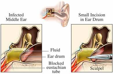
The middle ear is behind the eardrum or tympanic membrane. It is the space that houses the ear ossicles, the bones which transmit sound from the eardrum to the cochlea of the inner ear. This area is connected to the back of the nose (pharynx) via a hollow tube known as the eustachian tube.  An infection of this space is known as otitis media (OM). There are different types of OM, depending on the length of time it has persisted and whether or not fluid (effusion) is present.

Acute otitis media (AOM) typically occurs in <2 years old, usually caused by viruses such as RSV, cytomegalovirus (CMV) and Haemophilus influenzae (Hflu) but can also be due to bacteria. Mucosal congestion caused by the upper respiratory infection (URI) is aspirated up into the middle ear where inflammation occurs.
Treatment: If younger than 2 years old, an antibiotic such as amoxicillin will be prescribed. If older than 2 years old, doctors may choose to wait 48 hours to see if symptoms improve on their own before deciding to add an antibiotic. This is because studies have shown spontaneous resolution (without treatment) in up to 80% of this age group in 2-14 days.

Otitis media with effusion (OME) occurs when bacteria gets into the middle ear and fills it with pus. In growing children less than 7, the eustachian tube is often short and less vertical, so pus cannot drain properly and begins to back up. This growing pocket of fluid causes a large amount of pressure and pain to the child. The doctor will use a pneumatic otoscope to blow a burst of air at this ear drum. Normally the ear drum will move but in the case of OME, no movement occurs. Tympanometry may also be used to determine this. Long term OME (>3 episodes in 6 months or >4 episdoes in 12 months) may lead to hearing and language problems, so treatment is often indicated.
Treatment: myringotomy (hole is cut into the ear drum) with tympanostomy tube insertion is surgically performed. This allows fluid to freely drain out of the ear instead of backing up. It also alows antibiotic drops to be placed in the ear, which move through the tube and into the middle ear space to treat bacterial infections. Parents are instructed to apply the drops for 10 days whenever they see pus draining from the ears, which allows them to treat infections at home.


Reference
American Academy of Family Physicians, American Academy of Otolaryngology-Head and Neck Surgery, American Academy of Pediatrics Subcommittee on Otitis Media with Effusion. Otitis media with effusion. Pediatrics 2004; 113(5): 1412-29.